

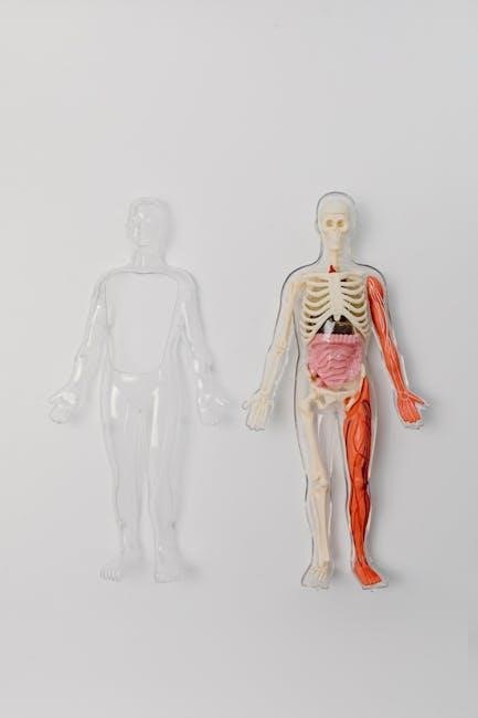
Grant’s Atlas, a cornerstone in anatomical education, provides detailed visuals. The 2017, 14th edition, and its
PDF availability are frequently sought by students and professionals alike, alongside resources like Gray’s and Chaurasia’s.
Overview of the Atlas
Grant’s Atlas of Anatomy stands as a premier resource for students and medical professionals seeking a comprehensive visual understanding of the human body. Renowned for its clarity and accuracy, the atlas presents anatomical structures through detailed illustrations and clinical imaging. The latest editions, particularly the 14th edition (2017), integrate modern radiological techniques – CT scans, MRIs, and more – alongside traditional dissection-based artwork.

Accessibility is key, with many seeking a Grant’s Atlas of Anatomy PDF version for convenient study. However, users should be aware of legitimate sources and potential risks associated with unauthorized downloads. The atlas’s organization follows a regional approach, facilitating focused learning. It’s often compared to other leading atlases like Gray’s and Netter’s, each offering unique strengths.

Historical Context and Authorship (Agur & Dalley)
Grant’s Atlas of Anatomy traces its origins back to J.C. Boileau Grant, initially published in the mid-20th century. However, the atlas achieved its current prominence under the stewardship of Arthur F. Dalley and A.J. Agur. Their collaborative work, beginning in the late 20th century, significantly refined the illustrations and expanded the clinical relevance of the atlas.
Agur and Dalley’s dedication to integrating modern imaging – a crucial aspect of the latest editions, including the sought-after Grant’s Atlas of Anatomy PDF – cemented its position as a leading educational tool. The 14th edition (2017) represents a culmination of their efforts, offering students a dynamic resource alongside texts like Gray’s and Chaurasia’s. Their commitment to accuracy and clarity continues to influence anatomical education.
Significance in Anatomical Education
Grant’s Atlas of Anatomy holds a pivotal role in medical, allied health, and biological sciences education; Its detailed, color-coded illustrations facilitate a deeper understanding of complex anatomical structures, complementing dissection labs and clinical studies. The availability of the Grant’s Atlas of Anatomy PDF further enhances accessibility for students globally.
Compared to alternatives like Gray’s and Netter’s, Grant’s emphasizes a clinically oriented approach, bridging the gap between theoretical knowledge and practical application. This focus, coupled with its regional organization, makes it invaluable for exam preparation and understanding patient cases. Its consistent updates, like the 2017 edition, ensure students learn from the most current anatomical understanding, solidifying its educational significance.
Key Features of the Latest Edition
Grant’s Atlas’s latest edition integrates advanced radiology, clinical correlations, and improved organization. The readily available PDF version enhances study convenience.
Enhanced Imaging Techniques (Radiology Integration)
Grant’s Atlas of Anatomy significantly elevates its visual learning experience through the seamless integration of modern radiological imaging. The latest edition expertly correlates anatomical structures with corresponding CT scans, MRIs, and other diagnostic imaging modalities. This innovative approach bridges the gap between traditional dissection-based anatomy and real-world clinical practice, allowing students to visualize anatomical relationships in vivo.
The inclusion of high-quality radiological images alongside detailed illustrations provides a more comprehensive understanding of anatomical structures and their clinical relevance. Students can now readily compare and contrast anatomical landmarks observed during dissection with their appearance on commonly used imaging techniques. Accessing the PDF version facilitates convenient study and review of these crucial imaging correlations, preparing future healthcare professionals for accurate diagnosis and treatment planning.
Clinical Correlations and Surface Anatomy
Grant’s Atlas of Anatomy excels in linking anatomical knowledge to clinical practice, a feature greatly enhanced in recent editions and readily accessible within the PDF format. Each anatomical region is accompanied by clinical boxes detailing relevant pathologies, common injuries, and surgical approaches. This contextualization transforms static anatomical study into a dynamic understanding of how structures function – and malfunction – in the human body.
Furthermore, the atlas emphasizes surface anatomy, illustrating palpable landmarks and their underlying structures. This is crucial for physical examinations and procedural guidance. The latest edition’s improved illustrations and accompanying text clearly demonstrate how anatomical knowledge translates to bedside skills. Students utilizing the digital PDF can easily cross-reference these clinical and surface anatomy details, solidifying their understanding and preparing them for clinical rotations.
Improved Organization and Regional Approach
Grant’s Atlas of Anatomy consistently refines its organizational structure, with the latest edition – often accessed as a convenient PDF – adopting a distinctly regional approach. This method groups structures by anatomical area (e.g., back, thorax, upper limb), mirroring how clinicians encounter anatomy in practice. This contrasts with systems-based approaches, fostering a more holistic and clinically relevant understanding.
The PDF version facilitates seamless navigation between regions and structures. Improved indexing and cross-referencing allow students to quickly locate specific information. The atlas’s layout prioritizes clarity, with consistent color-coding and labeling. This enhanced organization, readily available in the digital PDF format, streamlines the learning process and promotes efficient study, making complex anatomical relationships more accessible.

Regional Anatomy Coverage
Grant’s Atlas meticulously details anatomy by region – back, thorax, limbs – presented with clarity in the latest PDF edition for comprehensive study.
Back and Vertebral Column
Grant’s Atlas of Anatomy provides exceptionally detailed illustrations of the back and vertebral column, crucial for understanding musculoskeletal relationships and potential clinical presentations. The latest edition, often accessed as a PDF, showcases intricate depictions of vertebrae, ligaments, and associated musculature.
Students benefit from clear visualizations of spinal curves, intervertebral discs, and the complex network of nerves traversing this region. The atlas expertly illustrates the bony landmarks and surface anatomy, aiding in palpation and clinical examination skills. Furthermore, the PDF version facilitates easy navigation and focused study of specific vertebral levels and associated structures. This regional coverage, combined with clinical correlations, makes Grant’s an invaluable resource for medical and allied health professionals.
Thorax
Grant’s Atlas of Anatomy excels in its detailed depiction of the thorax, a region vital for respiration and protection of vital organs. The latest edition, frequently available as a convenient PDF, offers layered illustrations revealing the skeletal framework – ribs, sternum, and thoracic vertebrae – alongside the intercostal muscles and neurovascular bundles.
The atlas clearly demonstrates the position of the lungs, heart, and major vessels within the thoracic cavity. Students utilizing the PDF format can easily zoom and study the intricate relationships between these structures. Clinical correlations highlight potential sites of injury and disease. Grant’s provides a comprehensive understanding of surface anatomy, aiding in auscultation and percussion techniques, making it a crucial resource for medical education and practice.
Upper Limb
Grant’s Atlas of Anatomy provides exceptionally detailed illustrations of the upper limb, crucial for understanding its complex functionality. The latest edition, often accessed as a PDF, meticulously showcases the skeletal structure – clavicle, scapula, humerus, radius, and ulna – alongside associated ligaments and joints. Layered muscle depictions reveal origins, insertions, and actions, vital for clinical application.
The PDF version allows for focused study of neurovascular pathways, including brachial plexus branches and major arteries/veins. Grant’s excels in demonstrating the intricate relationship between structures within compartments of the arm and forearm. Clinical boxes correlate anatomical knowledge with common injuries and conditions. This comprehensive coverage makes it an invaluable resource for students and practitioners alike.
Lower Limb
Grant’s Atlas of Anatomy delivers a thorough visual guide to the lower limb, frequently utilized in conjunction with texts like Gray’s Anatomy for Students. The latest edition, often found as a convenient PDF, meticulously details the pelvic girdle, femur, tibia, fibula, and foot. Clear illustrations showcase ligaments, tendons, and joint articulations, essential for understanding biomechanics.
The PDF format facilitates focused study of muscle origins, insertions, and nerve innervation, crucial for diagnosing and treating lower limb pathologies. Grant’s excels in depicting fascial compartments and vascular supply. Clinical correlations within the atlas link anatomical structures to common injuries like ACL tears and compartment syndrome, enhancing practical understanding for medical professionals and students.

Neuroanatomy Section
Grant’s Atlas provides detailed brain, spinal cord, and nerve illustrations. The latest edition PDF offers clear depictions aiding neurological study and clinical correlation.
Brain Dissection and Imaging
Grant’s Atlas of Anatomy excels in its neuroanatomy section, particularly regarding brain visualization. The latest edition PDF incorporates advanced imaging techniques, seamlessly integrating radiological views with traditional dissection illustrations. This allows students to correlate anatomical structures observed during dissection with corresponding findings on CT scans and MRI images.
Detailed depictions of gyri, sulci, and functional areas are presented, enhancing understanding of cortical organization. The atlas provides comprehensive coverage of deep brain structures, including the basal ganglia, thalamus, and hypothalamus. Furthermore, cross-sectional views aid in spatial comprehension, crucial for interpreting clinical imaging. Accessing the PDF version facilitates convenient study and review, complementing hands-on dissection experiences and solidifying neurological knowledge.
Spinal Cord and Peripheral Nerves
Grant’s Atlas of Anatomy delivers exceptional detail in its depiction of the central and peripheral nervous systems. The latest edition PDF offers meticulously illustrated cross-sections of the spinal cord at various levels, clearly demonstrating the organization of gray and white matter, and the location of ascending and descending tracts.
Peripheral nerve distributions are comprehensively mapped, with accompanying surface anatomy illustrations aiding in clinical correlation. The atlas showcases nerve pathways and plexuses, crucial for understanding innervation patterns. Access to the PDF version allows for focused study of these complex structures, enhancing comprehension of neurological function and potential injury sites. This detailed presentation, combined with imaging integration, makes Grant’s an invaluable resource.

Cranial Nerves – Detailed Illustrations
Grant’s Atlas of Anatomy excels in its visualization of the cranial nerves, a notoriously challenging area for students. The latest edition PDF provides exceptionally clear illustrations detailing the course and branching patterns of each nerve. These depictions are paired with accompanying tables summarizing function – sensory, motor, or mixed – and clinical correlations.
Diagrams showcase the nuclei within the brainstem responsible for each nerve, alongside representations of their pathways. The PDF format facilitates close examination of these intricate details, aiding in memorization and understanding. Grant’s integrates radiological imaging to demonstrate nerve visualization in vivo, bridging anatomical study with clinical practice. This comprehensive approach solidifies understanding of cranial nerve function and dysfunction.

Accessing the PDF Version
Grant’s Atlas’s latest edition PDF is available through legitimate academic sources, but unauthorized downloads pose risks regarding copyright and file integrity.
Legitimate Sources for PDF Download
Accessing Grant’s Atlas of Anatomy in PDF format requires careful consideration of source legitimacy. Officially, the publisher, Elsevier, offers the atlas through their online bookstore and subscription services like ClinicalKey. University libraries frequently provide access to digital versions for enrolled students and faculty, ensuring authorized use.
Furthermore, some educational platforms and learning management systems may integrate the PDF directly into course materials. Checking with your institution’s library or course instructor is a reliable way to confirm access. Beware of websites offering “free” downloads, as these often violate copyright and may contain malware or outdated content. Prioritizing official channels guarantees a safe and legally compliant experience with this essential anatomical resource.
Potential Risks of Unauthorized Downloads
Downloading Grant’s Atlas of Anatomy from unofficial sources carries significant risks. These PDFs often harbor malware, viruses, and spyware, compromising your device’s security and potentially stealing personal information. Beyond technical threats, unauthorized downloads constitute copyright infringement, a legal offense with potential penalties.
Furthermore, illegally obtained PDFs may be incomplete, contain errors, or represent outdated editions, hindering accurate anatomical study. The quality of images can be severely compromised, making detailed observation difficult. Supporting legitimate sources ensures access to a reliable, updated, and secure resource. Protecting your digital wellbeing and respecting intellectual property rights are crucial when seeking anatomical learning materials.
Copyright and Ethical Considerations
Grant’s Atlas of Anatomy is protected by copyright law, safeguarding the intellectual property of its authors (Agur & Dalley) and publishers. Unauthorized distribution or reproduction of the PDF version is a violation of these rights, impacting the creators’ livelihood and future work.
Ethically, accessing anatomical resources through legitimate channels demonstrates respect for the effort and expertise invested in their creation. Utilizing legally obtained materials supports continued development and improvement of essential educational tools. Supporting publishers ensures the availability of high-quality anatomical atlases for future generations of students and medical professionals. Respecting copyright fosters a culture of academic integrity.

Comparison with Other Anatomy Atlases
Grant’s Atlas distinguishes itself through clinical correlations, while Gray’s offers comprehensive detail, and Netter’s excels in artistic clarity, each serving unique learning styles.
Grant’s vs. Gray’s Anatomy
Grant’s Atlas of Anatomy and Gray’s Anatomy are both highly respected resources, yet cater to different learning preferences. Gray’s, traditionally, provides an exhaustive textual description alongside illustrations, delving deeply into anatomical intricacies. It’s a comprehensive textbook suitable for those desiring a thorough understanding of anatomical principles.
Conversely, Grant’s prioritizes visual learning, presenting anatomy through meticulously detailed and clinically-focused illustrations. While Gray’s is often favored for its depth, Grant’s excels in spatial understanding and applying anatomical knowledge to clinical scenarios. The latest editions of both continue to integrate radiological imaging, but Grant’s maintains a stronger emphasis on surface anatomy and its clinical relevance, making it a popular choice for medical and allied health students.
Grant’s vs. Netter’s Atlas of Human Anatomy
Grant’s Atlas of Anatomy and Netter’s Atlas of Human Anatomy are both premier visual resources, but differ in artistic style and emphasis. Netter’s is renowned for its beautifully rendered, artistic illustrations that prioritize clarity and aesthetic appeal, often employing a more stylized approach. This makes complex structures readily accessible, particularly for initial learning.
Grant’s, however, utilizes realistic dissections and cadaveric images, offering a more true-to-life representation of anatomical structures. While Netter’s excels in providing a broad overview, Grant’s often provides greater detail in specific regions, especially concerning clinical correlations and surface anatomy. Both integrate modern imaging techniques, but Grant’s leans towards a more clinically-oriented presentation, making it valuable for understanding applied anatomy.
Choosing the Right Atlas for Your Needs
Selecting the ideal anatomy atlas depends heavily on your learning style and course requirements. If you prefer artistic, easily digestible visuals, Netter’s Atlas might be preferable. However, if a realistic, dissection-based approach aligns with your learning, Grant’s Atlas is a strong contender.
Consider whether clinical correlations are crucial; Grant’s excels in this area. Students benefiting from integrated radiology will find both atlases useful, but Grant’s increasingly emphasizes this aspect. Access to a PDF version, while convenient, necessitates verifying legitimate sources to avoid copyright infringement and malware risks. Ultimately, reviewing sample images from both atlases and aligning them with your curriculum is paramount.

Utilizing Grant’s Atlas for Effective Study
Grant’s Atlas enhances learning through dissection labs and exam prep. Its PDF format offers convenient access, complementing coursework and fostering deeper anatomical understanding.
Integration with Dissection Labs
Grant’s Atlas of Anatomy serves as an invaluable companion during dissection labs, bridging the gap between theoretical knowledge and practical application. The detailed, clinically-focused illustrations allow students to confidently identify structures within cadavers, enhancing their spatial understanding.
Utilizing the PDF version facilitates seamless navigation between the lab bench and the atlas, enabling quick reference and verification of anatomical relationships. Students can cross-reference dissected specimens with the atlas’s precise depictions, solidifying their comprehension. The atlas’s regional approach mirrors typical dissection sequences, further streamlining the learning process.
Furthermore, the integration of surface anatomy within Grant’s aids in correlating internal structures with external landmarks, a crucial skill for clinical practice. This synergy between dissection and atlas study fosters a robust and lasting grasp of human anatomy.
Using the Atlas for Exam Preparation
Grant’s Atlas of Anatomy, particularly the readily accessible PDF version, is a powerful tool for exam preparation. Its high-quality illustrations and clear labeling facilitate efficient memorization of anatomical structures and their relationships. The atlas’s clinical correlations provide context, aiding in understanding the functional significance of anatomical features – often tested concepts.
Students can utilize the atlas for self-testing, covering up labels and attempting to identify structures from memory. The regional organization allows for focused study of specific body areas, aligning with typical exam formats.
Combined with resources like Gray’s and Chaurasia’s, Grant’s offers a comprehensive visual review. The PDF format enables convenient access on various devices, maximizing study time and reinforcing anatomical knowledge for successful exam outcomes.
Online Resources and Companion Website
Grant’s Atlas of Anatomy is often complemented by a robust online platform, enhancing the learning experience beyond the printed pages or PDF version. These digital resources typically include self-assessment questions, interactive quizzes, and additional clinical cases to test comprehension. The companion website frequently features labeled images for self-testing, reinforcing anatomical recall.
Access to supplementary materials, such as videos demonstrating dissection techniques or radiological imaging interpretations, can significantly deepen understanding. Students can leverage these tools to bridge the gap between theoretical knowledge and practical application.
Furthermore, online forums and communities allow for peer-to-peer learning and discussion, fostering a collaborative study environment. Utilizing these resources alongside the Grant’s PDF maximizes learning potential.