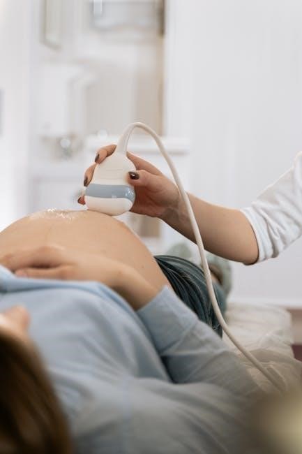
Overview of Ultrasound-Guided Breast Biopsy
Ultrasound-guided breast biopsy is a reliable method for obtaining tissue samples to determine if a lump is benign or malignant. It is faster, avoids radiation, and is often preferred for specific cases, such as in pregnant or lactating women.

1.1 Definition and Purpose
Ultrasound-guided breast biopsy is a minimally invasive procedure using ultrasound imaging to guide a needle for tissue sampling. It helps diagnose whether a breast lump is benign or malignant. The purpose is to evaluate suspicious lesions identified on imaging, providing accurate histologic assessment. This method is preferred for its precision, minimal invasiveness, and ability to target lesions that are difficult to access. It ensures timely diagnosis and appropriate treatment planning for patients.
1.2 Comparison with Other Biopsy Methods
Ultrasound-guided breast biopsy stands out for its real-time imaging, allowing precise needle placement without ionizing radiation. Unlike stereotactic or MRI-guided biopsies, it is faster and more cost-effective. Additionally, it avoids the discomfort of breast compression used in mammography-guided procedures. This method is particularly advantageous for superficial lesions and those near sensitive areas, making it a preferred choice for both patients and radiologists due to its efficiency and comfort.

CPT Codes for Ultrasound-Guided Breast Biopsy

Key CPT codes for ultrasound-guided breast biopsy include 76642 for imaging guidance and 77012 for diagnostic ultrasound. These codes are essential for accurate billing and documentation.
2.1 Primary CPT Codes Used
The primary CPT codes for ultrasound-guided breast biopsy include 76642 for ultrasound guidance and 77012 for diagnostic ultrasound imaging. Code 76642 covers the imaging guidance portion, while 77012 is used for the diagnostic ultrasound examination. These codes are essential for accurate billing and ensure proper reimbursement for the procedure. They specifically address the ultrasound components, distinguishing them from other biopsy-related codes. Proper coding is critical for compliance and streamlined insurance claims processing.
2.2 Additional Codes for Related Procedures

Additional CPT codes for ultrasound-guided breast biopsy include 10021 for fine-needle aspiration and 10120 for incisional biopsy. Codes like 10021 are used for aspirations, while 10120 applies to more invasive procedures. These codes complement the primary codes, ensuring comprehensive billing for all aspects of the biopsy. They cover various biopsy methods and tissue sampling approaches, ensuring accurate documentation and reimbursement for related services. Proper use of these codes is essential for complete claim submissions.

Indications for Ultrasound-Guided Breast Biopsy
Ultrasound-guided breast biopsy is indicated for suspicious lesions identified on ultrasound, such as solid masses or abnormalities requiring tissue sampling. It is also preferred for challenging cases, like lumps near the chest wall or under the arm, where other biopsy methods are less effective.
3.1 Suspicious Breast Lesions Identified on Ultrasound
Suspicious breast lesions detected via ultrasound, such as solid masses, irregular shapes, or non-circumscribed margins, often prompt a biopsy. These characteristics suggest potential malignancy, requiring tissue sampling for diagnosis. Ultrasound guidance ensures precise needle placement, minimizing complications. It’s particularly useful for lesions that are palpable or located near the chest wall, nipple, or implants, where other methods may struggle. This approach provides accurate histopathological assessment, crucial for timely treatment planning.
3.2 Challenges in Performing the Biopsy
Challenges include lesions near the nipple, skin, or implants, small or posterior targets in dense tissue, and mobile masses. These require modified techniques, such as open-trough or tandem-needle methods, to ensure accurate sampling while minimizing complications. Proper positioning and adjustments during the procedure are critical to overcome these difficulties and achieve diagnostic results.
Advantages of Ultrasound-Guided Breast Biopsy
Ultrasound-guided breast biopsy offers reduced radiation exposure, faster procedures, and cost-effectiveness. It allows for real-time imaging, improving accuracy and minimizing recovery time, making it a preferred option.
4.1 Reduced Radiation Exposure
Ultrasound-guided breast biopsy eliminates the need for ionizing radiation, making it safer for patients, especially pregnant or lactating women. Unlike stereotactic biopsies, it uses sound waves, reducing exposure risks while maintaining diagnostic accuracy. This method is preferred for its non-invasive approach and lower health risks compared to radiation-based procedures, ensuring patient safety without compromising results. It’s a significant advantage over other biopsy techniques.
4.2 Cost-Effectiveness and Faster Procedure
Ultrasound-guided breast biopsy is both cost-effective and efficient, reducing overall healthcare expenses. It is less expensive than stereotactic or surgical biopsies, with shorter procedure times and faster recovery. Patients can quickly resume daily activities, minimizing downtime. This method is preferred for its economic benefits and rapid results, making it a practical choice for patients and healthcare providers alike.
Devices and Techniques Used
Spring-loaded core needle biopsy devices and vacuum-assisted breast biopsy (VABB) devices are commonly used, offering precise tissue sampling with minimal complications.
5.1 Spring-Loaded Core Needle Biopsy Devices
Spring-loaded core needle biopsy devices use a two-step mechanism, deploying an inner needle to collect tissue samples, followed by an outer cannula to secure the specimen. These devices allow precise tissue sampling with minimal discomfort. They are commonly used for their reliability and ability to provide accurate diagnostic results, making them a preferred choice for ultrasound-guided breast biopsies.
5.2 Vacuum-Assisted Breast Biopsy (VABB) Devices
Vacuum-assisted breast biopsy devices enable efficient tissue sampling, especially for small or complex lesions. They use a vacuum to pull tissue into the device, allowing multiple samples with a single needle insertion. This method is ideal for lesions that are difficult to visualize or require comprehensive sampling, ensuring accurate diagnosis while minimizing patient discomfort and procedural trauma.
Step-by-Step Procedural Overview
The procedure begins with patient consent and preparation, followed by local anesthesia, skin cleaning, and ultrasound-guided needle insertion for tissue sampling, ensuring precise and efficient biopsy collection.
6.1 Patient Preparation and Positioning
Patient preparation involves obtaining consent, discussing risks, and positioning the patient to optimize breast tissue accessibility. Local anesthesia is administered, and the skin is cleaned with antiseptic solutions. The ultrasound transducer is disinfected or covered with a sterile sleeve. The patient is positioned to allow easy needle insertion, often with the affected breast exposed and supported. The procedural tray is prepared, including biopsy devices and anesthesia equipment, ensuring a sterile field for the procedure.
6.2 Needle Placement and Tissue Sampling
Needle placement is guided by ultrasound to ensure precise targeting of the lesion. Spring-loaded core needle biopsy devices deploy a sampling trough, followed by a cutting cannula to collect tissue. Vacuum-assisted devices allow multiple samples with a single insertion, improving accuracy. The procedure minimizes complications, ensuring adequate tissue for diagnosis while maintaining clear visualization of the needle throughout the process.

Post-Biopsy Care and Follow-Up
After the procedure, pressure is applied to the biopsy site, and a bandage is placed. Patients can resume normal activities quickly but should avoid strenuous exercise. Follow-up appointments are scheduled to review results and discuss further management.
7.1 Immediate Recovery and Wound Care
After the biopsy, pressure is applied to the site, and a bandage is placed. Patients can return home shortly and resume normal activities but should avoid strenuous exercise. The area may bruise or swell slightly. Keeping the wound dry and clean is essential to prevent infection. Over-the-counter pain relievers can manage discomfort. Complications are rare but may include bleeding or infection. Patients should contact their doctor if severe symptoms arise.
7.2 Interpretation of Biopsy Results
The biopsy results are analyzed by a pathologist to determine if the tissue is benign or malignant. A benign result indicates no cancer, while malignant means cancer cells are present. Atypical findings may require further testing. The results guide next steps, such as monitoring or treatment plans. Accurate interpretation is crucial for proper patient care and ensuring appropriate follow-up or therapy if cancer is diagnosed.

Coding and Billing Considerations
Accurate coding is essential for billing. CPT codes like 76942 (ultrasound guidance) and 19102 or 191025 (biopsy) ensure proper reimbursement and compliance with insurance requirements.
8.1 Reimbursement and Insurance Coverage
Reimbursement for ultrasound-guided breast biopsy varies by insurance. Most plans cover diagnostic biopsies when deemed medically necessary. CPT codes like 76942 (ultrasound guidance) and 19102 or 191025 (biopsy procedure) are typically used for billing. These codes ensure accurate reimbursement. However, coverage depends on the patient’s insurance plan and specific policy details. It is essential to verify coverage beforehand to avoid billing issues and ensure proper payment for the procedure.
8.2 Common Coding Errors to Avoid
Common coding errors include using incorrect CPT codes for ultrasound-guided breast biopsies. For example, using 77002 instead of 76942 can lead to denied claims. Mixing up biopsy types, such as stereotactic or MRI-guided, with ultrasound-guided procedures also causes errors. Additionally, failing to report both the biopsy and ultrasound guidance codes together may result in underpayment. Accurate and specific coding is crucial to ensure proper reimbursement and avoid billing delays or rejections.

Ultrasound-guided breast biopsy is a precise and efficient diagnostic tool, offering reduced radiation exposure and cost-effectiveness. Its accuracy and advancements continue to enhance patient care and outcomes significantly.
9.1 Summary of Key Points
Ultrasound-guided breast biopsy is a precise and efficient diagnostic method, offering reduced radiation exposure and cost-effectiveness. It is highly accurate for evaluating suspicious lesions and provides tissue samples for cancer diagnosis. The procedure is minimally invasive, with faster recovery times compared to surgical biopsies. Advances in techniques, such as spring-loaded core needles and vacuum-assisted devices, enhance diagnostic accuracy. This method is particularly beneficial for challenging cases, including deep or dense tissue lesions, ensuring optimal patient care and outcomes.
9.2 Future Directions in Ultrasound-Guided Breast Biopsy
Future advancements in ultrasound-guided breast biopsy may include improved imaging systems for better lesion visualization and real-time guidance. Innovations in biopsy devices, such as smaller needles or advanced vacuum-assisted systems, could enhance sampling accuracy. Techniques like hydrodissection may reduce complications in challenging cases. Additionally, integrating artificial intelligence could improve lesion detection and reduce human error, while combining ultrasound with other imaging modalities may offer comprehensive diagnostic solutions.

